Minimally invasive
Removes calcium deposits and scar tissue through a tiny skin puncture, no stitches, no large incisions.
Service
Targeted. Trusted. Minimally invasive.
Overview
At Mississippi Motion, we offer Tenex, a precise, minimally invasive procedure for patients with chronic tendon pain or calcific deposits that have not improved with rest, therapy, or injections.
Using ultrasound guidance and a specialized ultrasonic tool, Tenex removes only the damaged tissue or calcium buildup causing your pain, without harming healthy tissue or requiring open surgery.
The result? A same-day, in-office procedure with no stitches, no general anesthesia, and minimal downtime.
Philosophy of care
Tenex is built around a powerful concept: treat the root of the pain, not just the symptoms.
Instead of masking pain or repeatedly injecting tendons, Tenex:
Indications
Tenex is ideal for chronic tendon pain caused by overuse, injury, or scar tissue, and for calcific conditions that traditional treatments cannot resolve.
Common uses include:
If your tendon has been painful for over 3 months and shows thickening, disorganization, or calcium deposits on ultrasound, Tenex may be the next step.
Precise. Targeted. Same-day solution.
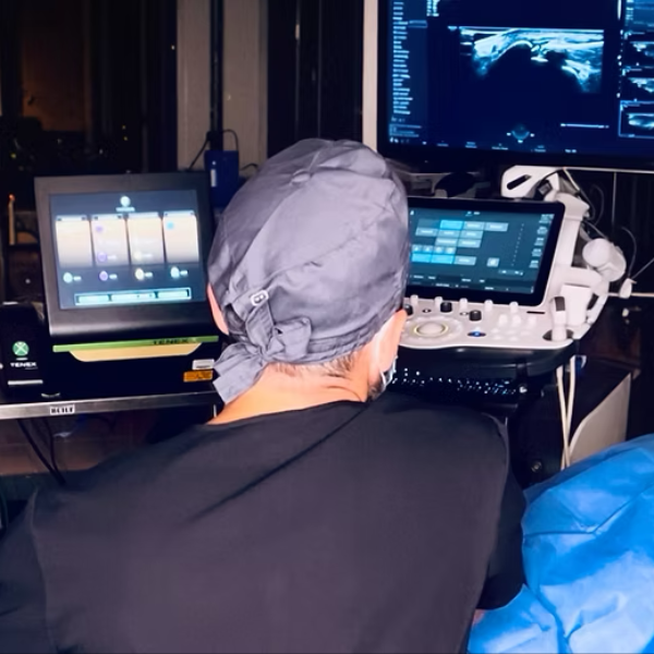
Step by step
We use diagnostic ultrasound to map the damaged area.
The skin is numbed, no sedation or general anesthesia required.
A tiny Tenex probe is placed through a small puncture site.
The ultrasonic tip vibrates at high frequency to break up and suction out damaged tendon tissue or calcium deposits, sparing healthy fibers.
A small bandage is applied, no stitches needed. Most patients walk out and resume light activity within a few days.
By region
| Region | Conditions treated |
|---|---|
| Elbow | Tennis elbow, golfer's elbow, elbow impingement |
| Shoulder | Rotator cuff tendinopathy (including calcific) |
| Knee | Patellar tendonitis, patellar osteophytes, see related video |
| Foot & heel | Plantar fasciitis, Achilles tendinopathy (calcific) |
| Hip | Gluteal or hamstring tendinopathy (incl. calcific) |
| Ankle | Insertional tendinopathy, ankle impingement |
Advanced technology. Real healing.
Video
Knee-oriented perspective, same technology highlighted in our conditions table above.
Why patients choose Tenex
Whether you are a runner, a weekend warrior, or simply tired of chronic pain, Tenex offers a precise and proven alternative to surgery.
Removes calcium deposits and scar tissue through a tiny skin puncture, no stitches, no large incisions.
Performed comfortably using local anesthetic or light sedation, without the need for intubation.
Most treatments take just 10–15 minutes, and patients resume light activity shortly after.
With minimal tissue disruption and healthy structures preserved, most patients resume light activity within days.
By removing the true source of pain, not just managing symptoms, Tenex supports lasting improvements in comfort and function.
If you are a candidate, Tenex may be performed within the same week of your evaluation.
Next step
We will assess your condition using diagnostic ultrasound, review your treatment options, and determine whether Tenex is a fit. We can often schedule the procedure within a week of your evaluation.
Choosing a procedure
Both TenJet and Tenex are minimally invasive options for chronic tendon conditions. They use different technologies and are optimized for different tissue types. The table below outlines key differences; Dr. Penton will recommend what fits your exam and imaging.
| Feature | Tenex (bone tip) | TenJet |
|---|---|---|
| Primary use | Removal of bony overgrowth and enthesophytes | Debridement of soft tissue tendinopathy, including insertional sites |
| Mechanism | Ultrasonic energy with hardened tip to break down fibrotic/calcific tissue at the bone–tendon interface | High-pressure dual-stream saline jet that dissects tissue and aspirates |
| Tissue types targeted | Scarred tendon, calcifications, enthesophytes, adherent tissue on bone | Chronic tendinosis, bursal tissue, fibrotic adhesions |
| Tip design | Hard, metallic beveled tip to gently shave tissue near bone | Dual-lumen catheter with angled fluid jet ports |
| Debridement control | High precision, especially at the osteotendinous junction | Broader debridement field |
| Ultrasound visibility | High, metallic tip is clearly visible under ultrasound | High, probe and jet activity are tracked in real time |
| Tissue removal efficiency | High for dense, calcific or bony lesions | High for diffuse or soft fibrotic tissue |
| Noise level during use | Moderate ultrasonic hum | Noticeable jet/water noise |
| Cleared for use (trained physician) | Cleared for use near bone/tendon junction | Cleared for soft tissue and fascia |
| Best clinical cases | Insertional Achilles with calcific changes, patellar tendinopathy with bony spur, rotator cuff tendinosis | Mid-substance tendinopathy, gluteus medius/minimus tears, rotator cuff tendinosis |
Training & affiliations